|
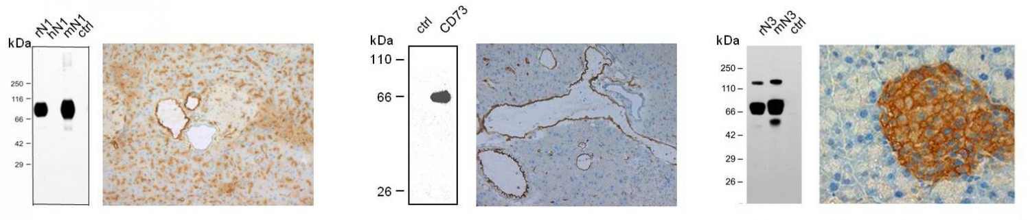
Western blot (left panel) and immunohistochemistry (right panel) with the guinea pig anti-mouse NTPDase1 antibody mN1-2cI5. Western blot shows specific immunoreactivity with bands corresponding to mouse (mN1) and rat (rN1) isoform present in cell lysates from COS transfected cells. Human NTPDase1 (hN1) is not detected by this antibody. Immunohistochemistry shows the presence of NTPDase1 in blood vessels (both EC and SMC), on the apical surface of ascini and in zymogen granules of a mouse pancreas. |
|
Western blot (left panel) and immunohistochemistry (right panel) with the rabbit anti-rat ecto-5'-Nucleotidase/CD73 rNu-9lI5 show immunoreactivity only in lysates from COS-7 cells transfected with a vector expressing rat CD73. The right panel shows the presence of this enzyme in blood vessels and capillaries of a mouse pancreas. |
|
Western blot (left panel) and immunohistochemistry (right panel) with the guinea pig anti-mouse NTPDase3 serum mN3-1cI4. The left panel shows specific immunoreactivity with mouse and rat NTPDase3 bands (dimer, monomer and a truncated fragment). The right panel shows NTPDase3 expression in Langerhans islet cells. |
|